
Cartilage Injury & Restoration in Boise
Sports Medicine and Physical Therapy Services for Knee Cartilage Damage in Boise.
What is a Cartilage Injury? A cartilage injury is damage to the smooth articular cartilage that lines the joint surfaces of the knee. These injuries range from small softening or fissuring of the surface to full-thickness defects that expose the underlying bone. Symptoms often include pain with activity, swelling, catching or locking, and a grinding sensation. Because articular cartilage has a very limited blood supply, it does not heal well on its own, which is why targeted treatment, and in some cases restoration surgery may be needed.
Understanding the Injury
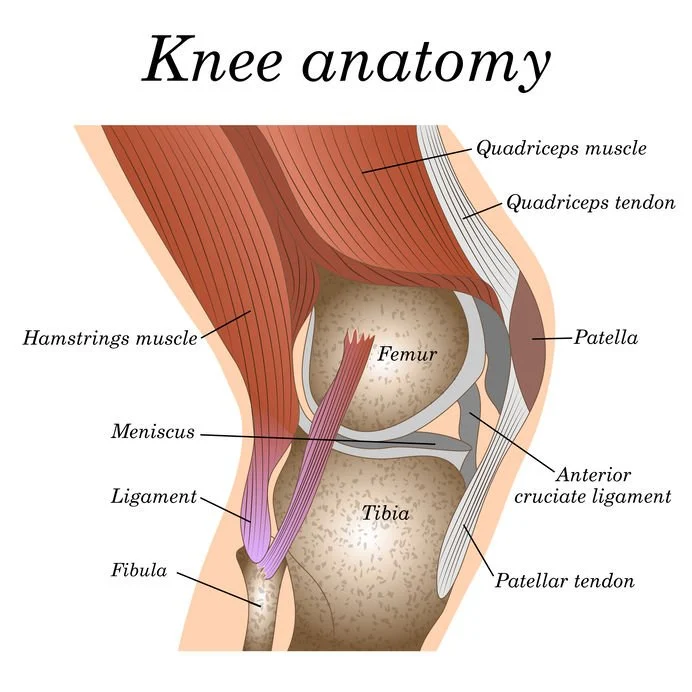
What Area of the Knee is Affected?
What Does the Exam Entail?
After discussing your history including sports and fitness activities, prior injuries, and any mechanical symptoms like catching or locking, your doctor will examine the knee. The exam assesses swelling (effusion), range of motion, joint-line tenderness, and quadriceps tone. Checking for crepitus (a rough, grinding feeling) as the knee bends and straightens can indicate articular cartilage breakdown, and specific tests help localize the affected compartment.
X-rays are used to evaluate joint alignment, joint-space narrowing, and to rule out other injuries, though cartilage itself is not visible on plain film. An MRI is the key imaging study for cartilage injuries: it can show the depth and size of a defect, the condition of the surrounding cartilage and bone, and any associated damage to the meniscus or ligaments. This information is essential for determining whether a defect can be treated conservatively or is a candidate for restoration.
Know the Injury
What are the Causes of Cartilage Injury?
Cartilage can be damaged suddenly by trauma or gradually through repetitive stress and joint overload. Primary contributors are acute impact injuries, joint instability, malalignment, and the wear associated with degeneration.
Acute Injury (Trauma)
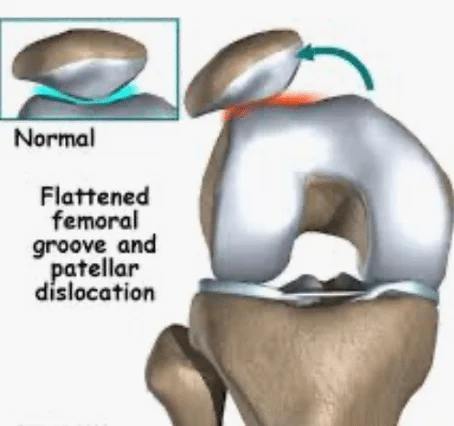
A direct blow, a twisting injury, or a joint dislocation can shear off a piece of cartilage or create a focal defect. These often occur alongside other injuries such as ACL tears or patellar dislocations. In younger patients, a fragment of cartilage and bone can separate from the surface, a condition known as an osteochondral lesion.
Joint Instability & Malalignment
When the knee is unstable—such as after an untreated ligament injury—or when the limb is malaligned, load is distributed unevenly across the joint surfaces. Concentrated stress on one area accelerates cartilage wear. Patellar maltracking, for example, can overload the cartilage on the underside of the kneecap.
Repetitive Overload & Degeneration
Repetitive high-impact activity, excess body weight, and prior meniscus loss all increase stress on the cartilage over time. As the surface softens and thins, small defects can enlarge and eventually progress toward osteoarthritis.
Meniscus Deficiency
The meniscus acts as a shock absorber and load distributor. When a significant portion of the meniscus is torn or removed, the cartilage bears more direct force, leaving it more vulnerable to damage.
What is the Treatment?
Treatment depends on the size, depth, and location of the defect, as well as your age, activity level, and goals. Many cartilage injuries are first managed conservatively with activity modification, physical therapy to strengthen the quadriceps, hips, and hamstrings, weight management, and anti-inflammatory measures. Correcting underlying issues such as instability, maltracking, or malalignment, is an important part of protecting the cartilage. Injections such as corticosteroids, viscosupplementation, or PRP may help manage symptoms depending on a person's short and long-term goals.
When conservative treatment is not enough, or when a defect is large or full-thickness, cartilage restoration surgery may be indicated. Common techniques include debridement (smoothing frayed cartilage), microfracture (creating tiny openings in the bone to stimulate a repair tissue response), osteochondral autograft or allograft transfer (transplanting cartilage-and-bone plugs), and cell-based approaches such as autologous chondrocyte implantation (ACI), which uses the patient's own cultured cartilage cells to fill a defect. In some cases, addressing alignment or instability at the same time gives the restored cartilage the best chance to succeed. Your doctor can discuss which options are most appropriate for you.
Need Help with Chronic or Recently Injured Cartilage Issue?
Schedule Your Appointment Today!